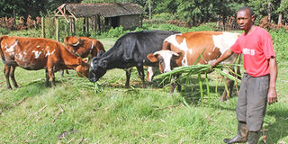
The caller on the line sounded anxious and kept pushing me to give him a definite answer. He complained I was asking too many questions while he had already sent me a photo of his sick cow.
I politely, but firmly reminded the caller, named Kamau, that I had already studied the photograph but I needed to know the start and progression of the problem, including any interventions that could have been taken at different times by him or his animal health service providers.
This is what we medically call the history of the case. It facilitates the doctor to know whether she is dealing with the initial problem or complications from other factors that may include attempts at treatment.
He calmed down. He told me he was a first-time subsistence dairy farmer and was not aware doctors needed a lot of information before identifying the problem.
We normally say veterinary doctors are like paediatricians as one must get the disease history from a third party since the patients cannot speak in a language we understand.
Kamau patiently narrated the cow’s history. It had calved down for the first time about three months earlier. The calf was in good health.
He had another older and horned cow in the same pen. The sick animal had been well but two weeks after calving, a small swelling appeared on the right side of the lower abdomen. Sometimes the swelling would be visible and other times it would disappear.
Kamau initially thought those were normal changes of the body, especially since the animal had been pregnant. He even wondered whether the cow could have retained a calf.
Within one month of appearing, the swelling increased to the size of a tennis ball and became a growing permanent feature on the cow’s skin.
By the time the farmer called his regular animal health service provider, the swelling was approaching the size of a small pumpkin and the tip appeared to be tilting downwards.
The service provider demonstrated to Kamau that all the contents of the swelling could be pushed back into the abdomen leaving a hole covered by the skin.
She demonstrated the hole by pushing her fist partly into the abdomen, with the skin still intact. The fist went into the body up to her wrist.
The service provider diagnosed hernia, which is a break in the body wall where internal organs such as fat, intestinal loops or the omentum protrude through the hole and rest under the skin.
This pushes the skin outward to form a swelling. The ometum is the towel-like tissue that wraps abdominal organs and holds abdominal fat. The service provider said the hernia was too big to be repaired and she recommended disposal of the animal for meat.
The farmer said he called me for a second opinion. I concurred with the first diagnosis and recommendation.
Considering the rate at which the swelling was growing, it was evident that the body wall muscles had been damaged and weakened.
They could not hold the internal pressure of the body organs caused by the abdominal weight, body motion and the movements created by digestive activities.
Like the cow udder, the cattle digestive system is another factory in the animal’s body. It produces a lot of gas, fluids and digestive waste as it extracts and builds nutrients from the feed eaten.
When there is hernia, the gas, fluids and digestive waste keep pushing the intestines and other tissues deeper into the hole in the body wall hence tearing more muscles and enlarging the swelling.
If the hernia is prolonged, the intestines may attach to the inner side of the skin and get damaged or twisted. This end up killing the cow due to intestinal strangulation.
Small hernias can be surgically corrected as the doctor pushes the organs back into the body from the skin. She then opens the skin, identifies the torn muscles and stitches the body wall with strong suture material.
Attempting to repair large tears is futile because the hernias tend to recur or get infected due to the heavily weakened body wall.
I advised Kamau the hernia in his cow could have been caused by horn injury inflicted by the other cow. I advise farmers to dehorn all their animals, since horns are weapons of defence and offence.
Horned cows normally attack the hornless in competition for food, space and dominance. They may also attack and injure or kill humans.
Hernias are common swellings on the bodies of cattle and other livestock. They occur under the skin in areas where there is a body cavity formed by layers of soft tissue. The most common are abdominal, umbilical, scrotal and inguinal hernias.
Kamau’s cow had an abdominal hernia, which are seen on the body wall, mainly lower abdomen or the sides. They are mostly caused by blunt injury to the body wall or infections that first form deep pus-filled cavities called abscesses.
Umbilical hernias are swellings on the area surrounding the umbilical stump. They are normally seen in calves and young stock.
Most are caused by genetic abnormalities. Animals with umbilical hernias should not be selected for breeding to prevent propagation of the genetic trait.
In pigs, hernia development is associated with boars. Thus, boars that produce piglets with high incidence of hernias should not be used for breeding.
Scrotal and inguinal hernias are mainly seen in pigs where intestinal loops get lodged in the scrotum or inguinal canal.
Such pigs are castrated and the inguinal canal is closed with sutures. The inguinal canal is a body channel that connects the scrotum with the abdomen.
The purpose of the canal is to allow the testicles to descend from the abdomen to the scrotum close the birth of the male offspring. Thereafter, the canal is supposed to close.
A doctor confirms a hernia by identifying a break in the body wall, finding the swelling contents soft and being able to push them back into the abdomen. Some hernias may get inflamed and become hard and impossible to push back into the abdomen.
Other swellings on the bodies of animals include abscesses, injured or inflamed tissues and tumours. There are also swellings due to allergic reactions, blood or serum accumulation after injury.